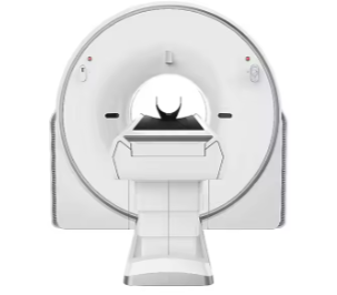
CT Scanner

Medical 16 Slice CT Scanner YSCT755

Model

YSCT755

Brand

YSENMED

Product Description

Optima Design


One Side Integrated Control

• Optimize System Control Layout. Improve Systematic Process Flow. Ensure Product Quality and Stability. Improve After-sales Maintenance Efficiency.


Thermal Insulation Design

• Improve Heat Dissipation Efficiency. Extend the Life of Detector. Ensure the Image Quality.


The Integrated Casting of Stator and Rotor

• Minimum Vibration During Rotation. Minimum Deformation During Rotation.


High Precision Bearing

• Zero Error and Zero Runout under High Speed Rotation. Achieve Military and Aerospace Level Requirements. Long Service Life and Excellent Stability.


Multi-point Temperature Control Technology

• Automatically Monitor the Temperature. Ensure the Stability of System Operation.


Clinical Application Image


Cloud diagnosis

• Famous radiologists diagnose through remote image diagnosis solution, improving primary hospital diagnosis ability.


Cloud storage

• MinFound Cloud storage is safe, stable and able to save much cost: payable based on requirement; it saves equipment purchasing and operation cost.


Global After-sales Service

• Automatic Fault Warning Function.

• Remote Service Function.


System Software

• Beam hardening artifact correction software.

• Posterior cranial fossa optimization.

• Motion correction reduces motion artifact.

• Pediatric protocol.

• NDI (NanoDose Iteration).

• Filming print software.

• Remote maintenance system.


Accessory KIT

• Patient Table Pad, Head Holder, Head Holder Cushion, Phantom.


Clinical Application

• Include Volume Reconstruction (VR), Multi-planar Reconstruction (MPR), Curved Planar Reconstruction (CPR), Surface Shaded Display (SSD), MIP, MinP.


Option

• UPS.

• Printer.

• High Pressure Injector.

• FOV: 500 mm.


Workstation

• 16GB Memory, 24″ color display, 1TB SATA hard disc.

• 3D image reconstruction: Include VR, MPR, CPR, SSD, Simulated scalpel, Virtual endoscope, CTA remove bone, CTA subtraction.

• Cerebral hemorrhage measuring tool.

• Skeleton internal fixation fluoroscopy technique.

• Advanced automatic melting of bone.

• CT vessel analysis.

• Bone fragment removal.

• ROI creator.

• Stent planning.

• Abdominal fat analysis.


Gantry
Aperture 70 cm
Manual control from POD at console Yes
Gantry cooling Air
Gantry rotation Belt-driven
Slip ring type Low voltage
Positioning system 3D lasers

HV Generator
Power rating 32 kW
mA range 10 – 300 mA
kV range 70 kV, 80 kV, 100 kV, 110 kV, 120 kV, 140

X-Ray tube
Anode heat storage capacity 5 MHU
Anode heat dissipation 735 KHU / мин
Small focal spot size 0.7 mm × 0.8
Large focal spot size 1.2 mm × 1.4

Detector
Number of detector rows 16
Number of detectors per row 704

Table
Maximum motion range 1600 mm
Speed in horizontal direction 150 mm/s
Motion tolerance in horizontal direction (200 kg) ±1mm
Table elevation range 440
Max weight load 450 lb (205 kg)
Foot pedal control, table horizontal motion Yes

Console
Uninterrupted power supply UPS 3 KVA to prevent sudden power outages and ensure the system stability
Memory 32 GВ
Hard disk 2TB + 1TB
Frequency 3.6 GHz
Liquid crystal display of high resolution, displaying matrix (M * M) 1920 × 1200
Progressive scan display Yes
DICOM 3.0 network interface Yes
Burn mode DICOM 3.0 print interface, DICOM 3.0 output and input interface, Auto-film
Auto-voice and integrated intercom For communication between operator and patient
DICOM Features Include DICOM PRINT, DICOM STORE, DICOM QUERY, DICOM RETRIVE
Interface Include HIS & RIS interface
Raw data reconstruction Raw data can be reconstructed on the workstation

Scan and Image Reconstruction
The fast rotation time 0.75 s / 360°
Longest scanning time 100 s
Longest scanning range 1200 mm on the original tabletop with head holder; 1300 mm on the original tabletop with the short extendet tabletop; 1400 mm on the original tabletop with the long extended tabletop
Scout scan direction Anteroposterior, lateral
Pitch range Standard 0.25 – 1.75. Optional: 0.1 – 2.0
Maximum number of slices per rotation 16 / 32 slices
Thinnest image slice thickness in 16-slice mode 25 mm
Thinnest image slice thickness in 32-slice mode 25 mm
Thinnest reconstructed slice thickness in 16-slice mode 25 mm
Thinnest reconstructed slice thickness in 32-slice mode 25 mm
Nominal slice thickness for axial scan 1.25 mm, 5 mm, 5 mm, 10 mm
Nominal slice thickness for helical scan 1.25 mm, 5 mm, 3.75 mm, 5.0 mm, 7.5 mm, 10 mm
Parallel image processing Yes
Simultaneous Reconstruction Can reconstruct and reorganize the images simultaneously with a variety of options. Different reconstruction options are available within the scan protocol
Scanning FOV
Image reconstruction matrix 1024 × 1024
Image display matrix 1024 × 1024

PDU
Input voltage 3-phase 380 V, 50 Hz
Power 50 kVA

Scroll to Top